We have all heard the phrase, “Your eyes are the window to your soul.” Medicine offers a more literal version of the idea, because the eyes also show the condition of the heart.
What Your Doctor Sees at the Back of Your Eye
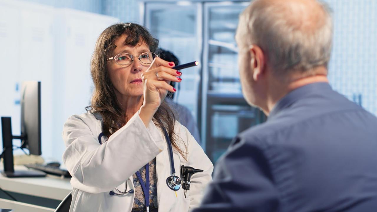
When an eye doctor shines a light into your pupil, they can see the retina, the lining at the back of the eyeball that is sensitive to light. The retina has a network of small arteries and veins across its surface. Many of them are narrower than a human hair. At other places in the body, small vessels like these lie under skin and muscle, and the retina is one of the few places where they can be seen and photographed.
High blood pressure, high blood sugar, smoking, alcohol consumption and age all leave visible marks on the vessels in the eye, such as narrowed or twisted vessels and leaks. These are the same kind of small vessels found in the heart and brain, and the same risk factors damage them all, so changes in the retina can indicate that similar damage is occurring in parts of the body that are harder to see or have not yet produced symptoms.
Why Early Detection Matters
Cardiovascular disease is among the leading causes of death taking over 19.8 million lives a year as per World Health Organization estimates. More than four in five of those deaths come from heart attacks and strokes, with one-third at ages below 70. India carries a heavy share with over 28% of deaths from cardiovascular disease.
The challenge in diagnosing cardiovascular disease early is that much of the damage happens without symptoms. About one heart attack in five is silent, with injury to the heart muscle even as the person remains unaware of the incident. A simple, painless test that flags risk early would help here, and the eye examination is a candidate.
What Blood Pressure, Blood Sugar, and Cholesterol Show in the Eye
High blood pressure affects the retina, with chronic hypertension leading to narrowing of the retinal blood vessels, a condition called hypertensive retinopathy. Thickened arteries can press on the veins where the two cross, a sign called arteriovenous nicking. In severe cases, small hemorrhages (bleeding) and fluffy white patches appear, indicating areas where there is less blood supply. Studies have shown that these signs are an early predictor of heart trouble. People with signs of retinopathy have double the risk of heart failure, and three times the risk among those with no existing heart disease, diabetes or high blood pressure.
High blood sugar damages the retinal vessels as well, in a condition called diabetic retinopathy, so people with diabetes have particular reason to keep up with eye examinations.
Some clues are also outside the retina. Xanthelasmata are soft, yellowish patches of cholesterol on the eyelids; arcus senili is a gray or white ring around the edge of the cornea. A study over three decades of adults showed that those who had xanthelasmata at the start, around 4% of the people being studied, went on to have more heart attacks, more severe atherosclerosis and higher death rates. Arcus, seen in about a quarter of participants, added little once other risk factors were counted, although in younger adults it can point to high cholesterol. Anyone who notices a yellowish patch on the eyelids or a ring around the cornea should ask a doctor for a cholesterol test.
When the Eye Signals an Emergency
The retina can also flag an emergency. A retinal artery occlusion happens when a clot or a fragment of plaque, most often from the carotid artery in the neck, blocks blood flow to the retina. The person loses vision in one eye, suddenly and without pain. This is the eye’s version of a stroke, with a high risk of stroke during the two weeks after the vision loss, and the risks of heart attack and death stay raised for more than a year.
Sudden loss of vision in one eye, even for a few minutes and even when vision returns, is a medical emergency. The person should go to a hospital with stroke care at once and should not wait for an appointment with a cardiologist, because a brief loss of vision can warn of a stroke, and the days that follow carry the highest risk.
What Artificial Intelligence Can Read From a Retinal Photograph
New research using artificial intelligence to read retinal photographs has produced encouraging results. Deep learning models can estimate a person’s age within about three years and their systolic blood pressure (the upper number in a reading) to within about 11 mmHg from a retinal photograph alone, and they can identify smokers with less accuracy. The same approach has been used to predict major cardiac events, with moderate accuracy.
A risk score built from age, sex, smoking, medical history and retinal vessel measurements performed as well as the Framingham risk score, a standard tool in clinics that estimates a person’s ten-year risk of heart disease, with no blood test or blood pressure reading needed. A second tool built on retinal photographs, called Reti-CVD, identified people at intermediate and high risk of cardiovascular disease in line with existing risk assessment tools.
What the Research Cannot Yet Show
Although retinal findings offer early information about heart health, they do not replace a blood pressure cuff or a cholesterol test. Trials are still required to show that screening people with retinal photographs leads to fewer heart attacks or strokes. Few studies have included South Asian populations, and more studies will be required to apply the findings to Indian patients.
What You Can Do
Regular eye examinations are worth keeping up. People with diabetes need a dilated eye examination, in which drops widen the pupil so the doctor can see the whole retina, at least once a year, and anyone with high blood pressure should ask an eye doctor how often to be checked. The doctor looks for narrowed vessels, bleeding and signs of hypertensive or diabetic retinopathy. Many eye hospitals and optical clinics offer retinal photography, and an eye doctor should review the images. The findings allow your physician to monitor and manage your blood pressure, cholesterol and blood sugar.
The same healthy habits that protect the heart protect the retina as well. Not smoking, avoiding alcohol consumption, keeping blood pressure and blood sugar under control, staying active and eating less salt all contribute to eye and heart health.
The retina is one of the few places where a doctor can look at small blood vessels of the kind found in the brain and heart, which turns a routine eye examination into a chance to catch vessel damage before it causes symptoms. What the doctor finds guides decisions about blood pressure, cholesterol, and blood sugar, and the vessels at the back of your eye show the results of keeping those in range.
Frequently Asked Questions
Can an eye examination detect heart disease?
An eye examination cannot diagnose heart disease, but it can show early damage to the small blood vessels of the retina, which often reflects similar damage in the heart and brain. Findings such as narrowed vessels or bleeding can prompt a doctor to check blood pressure, cholesterol, and blood sugar. The examination does not replace those tests.
What can the retina show about heart health?
The retina contains small arteries and veins that a doctor can see and photograph. High blood pressure, high blood sugar, smoking, alcohol consumption, and age leave visible marks on these vessels, including narrowing, twisting, and leaks. These marks can indicate similar damage in parts of the body that cannot be seen or have not yet produced symptoms.
What is hypertensive retinopathy?
Hypertensive retinopathy is damage to the retina caused by chronic high blood pressure. It begins with narrowing of the retinal blood vessels and, in severe cases, progresses to bleeding and fluffy white patches. Studies have linked these signs to a higher risk of heart failure and other heart trouble.
What do yellowish patches on the eyelids mean?
Yellowish patches on the eyelids, called xanthelasmata, are soft deposits of cholesterol. In a long-term study of adults, people who had xanthelasmata went on to have more heart attacks, more severe atherosclerosis, and higher death rates. Anyone who notices them should ask a doctor for a cholesterol test.
Is sudden vision loss in one eye an emergency?
Yes. Sudden vision loss in one eye, even for a few minutes and even when vision returns, can be a warning sign of stroke, and the days that follow carry the highest risk. The person should go to a hospital with stroke care at once and should not wait for an appointment with a cardiologist.
Can artificial intelligence assess heart risk from a retinal photograph?
Research suggests that artificial intelligence can contribute to heart risk assessment. Deep learning models have estimated age and systolic blood pressure from retinal photographs, and one risk score built on retinal vessel measurements performed as well as the Framingham score. Trials have yet to show that retinal screening leads to fewer heart attacks or strokes.
How often should I have my eyes examined?
People with diabetes need a dilated eye examination at least once a year, and people with high blood pressure should ask an eye doctor how often to be checked. In a dilated examination, drops widen the pupil so the doctor can see the whole retina and look for narrowed vessels, bleeding, and signs of retinopathy.
 Spot an error in this article? A typo maybe? Or an incorrect source? Let us know!